Category Archives: Orthopedics

Do Flip-flops with arch support work?

Foot orthotics really are a effective modality used by podiatric physicians to manage a wide range of foot problems. All the clinical experiences and research evidence is that they are very effective. Nevertheless, one trouble with them is that they have to be worn in footwear. That is naturally a lifestyle option, but sometimes the options and the environment do not necessarily accommodate the use of the right footwear which foot supports could be worn in.

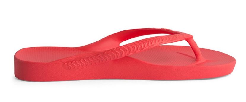
One query which you see asked frequently is that are those flip flops that come with an arch support built into them, can they be used instead of foot supports. There are a variety of manufacturers available on the market of flip flops that have different amounts of arch support built into them.

Are they as effective as foot supports?

That’s doubtful. The support that is included in them is just like what you will receive from a premade foot orthotics or one of the typical over-the-counter kind of foot supports. That is fine if you have an average arch shape. However, that is not good if you don’t. Foot orthotics usually are built to be specific to your foot type.

Should you use them?

There’s no harm in using these and they certainly might be used as an adjunct to foot supports when you’re not wearing footwear. As if they may be utilized as an alternative, you should discuss that with your foot doctor.

I do keep hearing about the Archies on the internet, however I haven’t seen them because they are from Australia. Evidently numerous podiatry clinics around Australia retail them.

Cuboid Syndrome

The cuboid is a smaller cube shaped bone on the lateral side of the foot around about the center of the foot. The bone is a bit bigger than a common gaming dice. The bone takes part in three joints and functions as a pulley for the tendon of the peroneus longus muscle to pass under. Because this is a powerful muscle it can move the cuboid bone too much if it is not steady and overload those joints that this bone is a part of producing a disorder known as cuboid syndrome. This is probably one of the more frequent causes of pain on the lateral side of the foot, particularly in athletes. The pain typically starts out quite mild and is located around where the cuboid bone is on the outside of the foot. The discomfort is only to begin with present during exercise. If the exercise levels are not lowered the problem will generally advance and then show up after exercise in addition to during. Occasionally the pain may radiate down into the foot. Although this is the commonest reason for pain here, there are others such as tendinopathy and nerve entrapments.

The main management of cuboid syndrome is pain relief. This is generally achieved with a decrease in exercise levels and the using of strapping to immobilise and support the cuboid. Mobilisation and manipulation is often used to fix the symptoms. Over the long run foot supports may be needed to control the movement and aid the lateral arch of the foot. This helps make the cuboid more stable so it is an efficient fulcrum or pulley for the tendon to work around. Generally this approach works in nearly all cases. If it doesn’t there are no surgical or more advanced methods and a further reduction in exercise levels is often the only alternative.

The Accessory Navicular

The accessory navicular is a supplementary bit of bone on the inside of the foot just on top of the mid-foot in the vicinity of its highest part. The bone is included within the tibialis posterior tendon that attaches to the navicular bone towards the top of the mid-foot ( arch ). The additional bone can also be known as the os navicularum or os tibiale externum. This is genetic, so is existing since birth. There are a few different kinds of accessory navicular and the Geist classification is most typically used. This classification divides the accessory navicular into 3 varieties:

Type 1 accessory navicular bone:
This is the classical ‘os tibiale externum’ and make up 30% of the occurrences; it is a 2-3mm sesamoid bone embedded inside the distal area of the tendon with no link to the navicular tuberosity and could be separated from it by up to 5mm

Type 2 accessory navicular bone:
This type makes up 55% of the accessory navicular bones; it’s triangular or heart-shaped and connected to the navicular bone through cartilage. It may well eventually join to the navicular to form one bone.

Type 3 accessory navicular bone:
Prominent navicular tuberosity. This could have been a Type 2 that has fused to the navicular

The typical symptom associated with an accessory navicular is the enlargement on the inside side of the mid-foot ( arch ). Because of the additional bone there, this impacts how well the mid-foot muscles do the job and may lead to a painful foot. Inflexible type shoes, like ice skates, may also be very uncomfortable to use because of the enlarged pronounced bone.

The treatment is geared towards the signs and symptoms. When the flatfoot is an issue, then ice, immobilisation and also pain relief medication may be required to start with. Following that, physical therapy and foot orthotic inserts to aid the foot are used. When the soreness is a result of pressure from the type of shoes which needs to be used, then donut type padding is required to get pressure off the painful region or the shoes might need to be modified.

If these non-surgical therapies fail to reduce the symptoms of the accessory navicular or maybe the issue is an ongoing one, then surgery may be a suitable option. This requires removing the accessory bone and restoring the insertion of the posterior tendon so its function is improved.